Compression and percussion tests
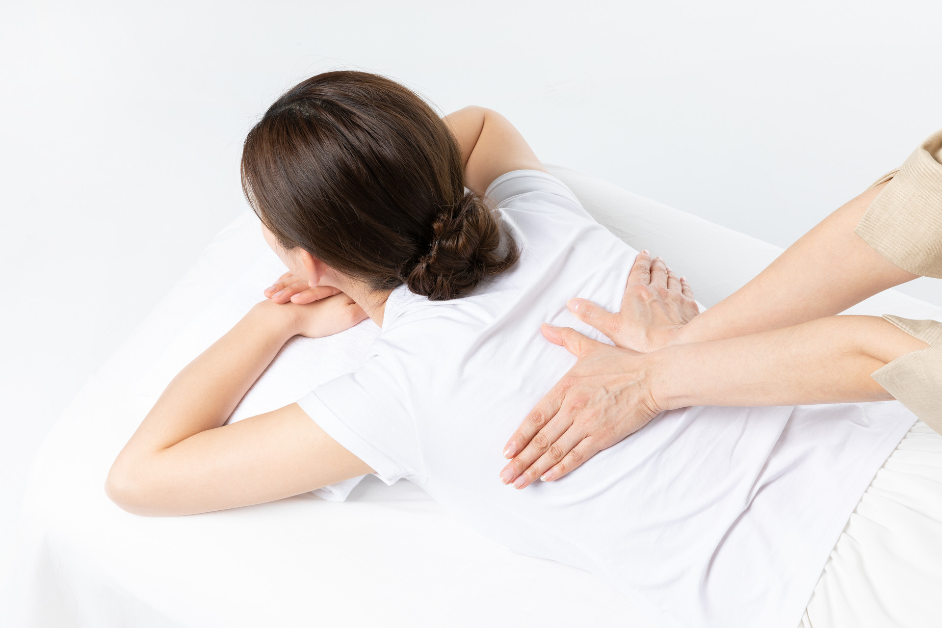
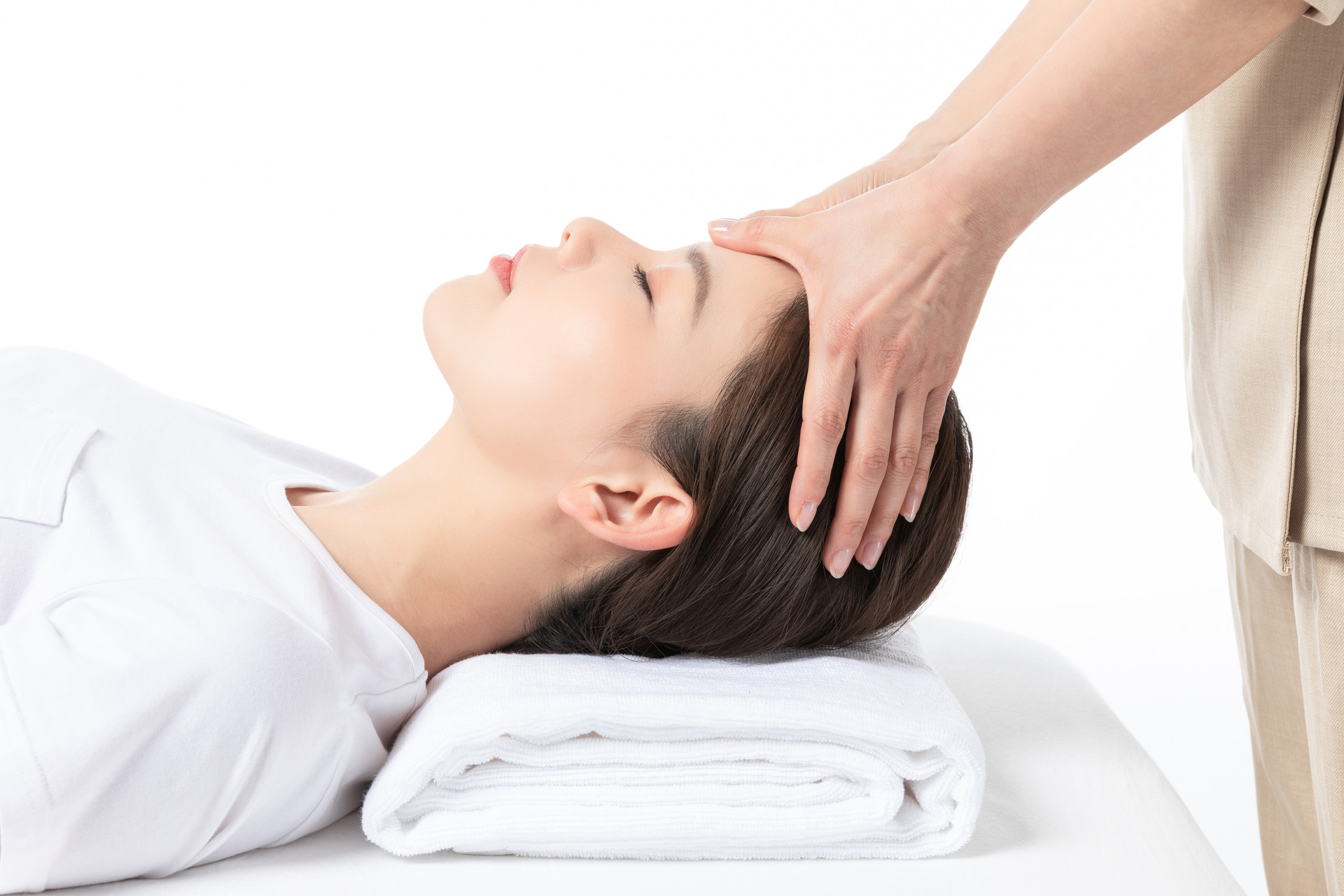
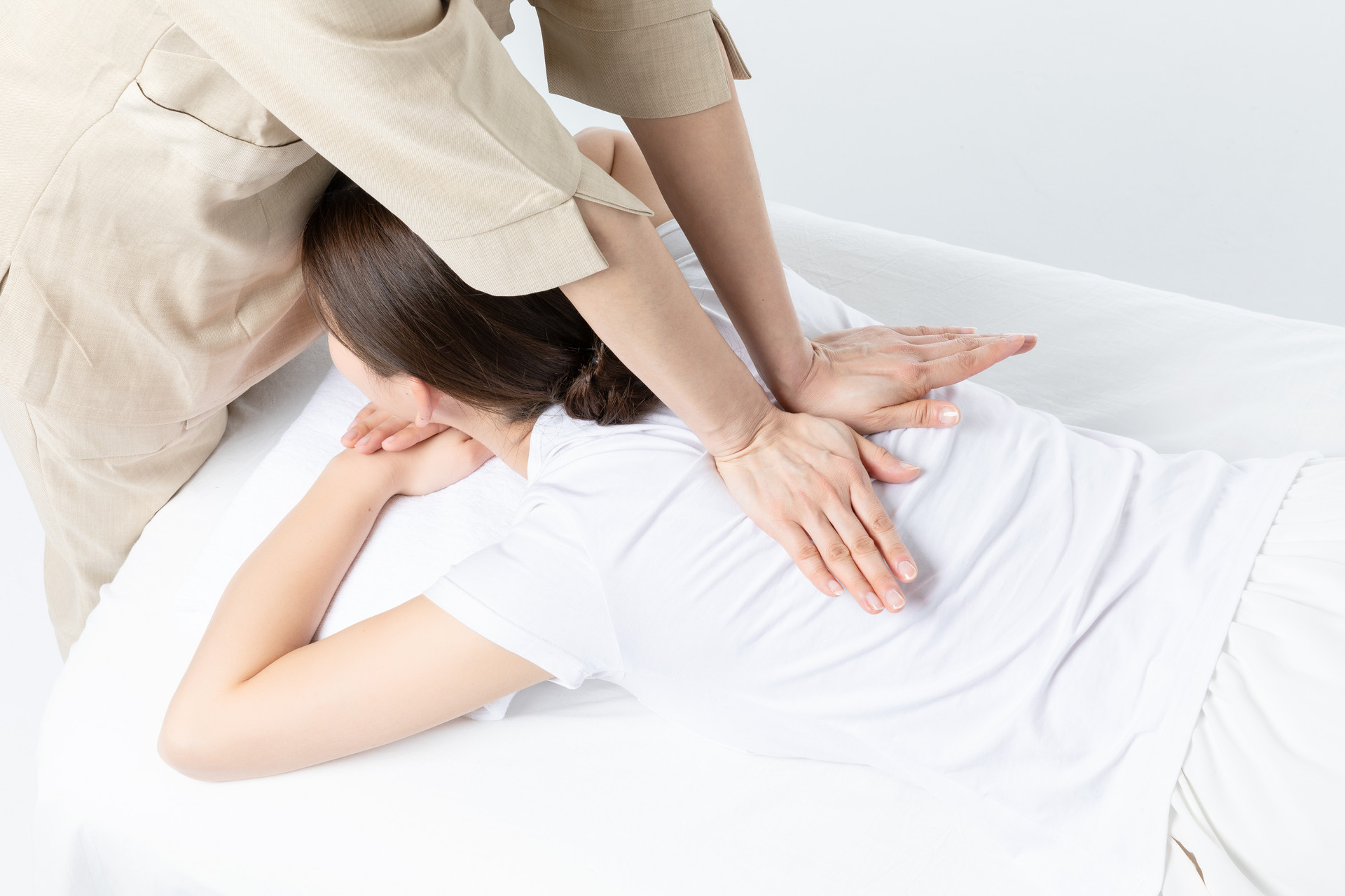
The patient sits while the doctor places their hands on top of the patient's head and applies pressure to different angles of the neck. If this causes neck pain and radiating pain, it indicates compression of the cervical nerve roots. In the upright position, the doctor uses a fist to tap the patient's head. If this causes neck pain and radiating pain or pain in the lower back and legs on the affected side, it is considered positive and suggests compression of the cervical or lumbar nerve roots.
Brachial plexus traction test
The patient sits with their neck flexed forward. The doctor stands on the affected side, with one hand supporting the patient's head and the other hand gripping the patient's wrist, pulling in the opposite direction. If this causes shooting pain or numbness in the affected limb, it indicates compression of the brachial plexus.
Head tilt test
The patient sits with their head slightly extended and their chin turned towards the affected side. They take a deep breath and hold it. The doctor applies resistance by pressing against the patient's chin with one hand, while the other hand feels the patient's radial artery. If the pulse weakens or disappears, it is considered positive and is often seen in scalene muscle syndrome.
Cervical traction test
The patient sits upright and relaxed. The doctor stands behind them, cupping their hands under the patient's occiput, and slowly lifts the patient's head. If the patient experiences relief from neck and shoulder pain and numbness, it is considered positive. This test is often used to determine if cervical traction is necessary.
Vertebral artery torsion test
The patient sits with their neck relaxed. The doctor stands behind the patient, holding their head with both hands to stabilize it, and asks the patient to perform maximal extension and rotation of the neck. If the patient experiences dizziness, vertigo, nausea, or vomiting, it is considered positive.
Neck flexion test
The patient sits with their legs extended and actively or passively flexes their neck, bringing their chin close to their chest for approximately one minute. If this causes lower back and leg pain, it is considered positive and suggests compression of the lumbar nerve roots.
Supine straight leg raise and dorsiflexion test
The patient lies flat on their back with both legs extended. While keeping the knee joint straight, the doctor lifts one leg at a time. The pain-free range of motion (angle between the lifted leg and the bed surface) is measured. If there is compression of the nerve roots, there will be a significant limitation in straight leg raise, usually below 60°, and pain in the distribution area of the compressed nerve roots, indicating a positive straight leg raise test. Then, the leg is lowered 5-10° until the pain disappears, and suddenly dorsiflexed. The reappearance of sciatic pain is considered positive and is more clinically valuable for diagnosing lumbar disc herniation compared to the previous test. It is important to note that a positive straight leg raise test can also occur in other lower limb conditions such as iliotibial band tension, while a positive dorsiflexion test is indicative of pure sciatic nerve tension.
Bedside test
The patient lies flat on their back with their affected side buttock close to the edge of the bed. The healthy side leg is flexed at the knee and hip joint, while the doctor holds the affected leg and extends it as far back as possible, causing distraction and movement of the sacroiliac joint. If there is pain in the sacroiliac joint, it indicates a lesion.
Heel to buttock test
The patient lies prone with both legs extended and relaxed. The doctor holds their foot and brings the heel to the buttock. If there is a lesion in the lumbosacral joint, it will cause pain in the lumbosacral region, and the pelvis and waist will also rise.
Dugas' test
In a normal person, when their hand is placed on the opposite shoulder, the elbow joint can touch the chest wall.
| 1 2 3 > >> >>|


